tree in bud nodules
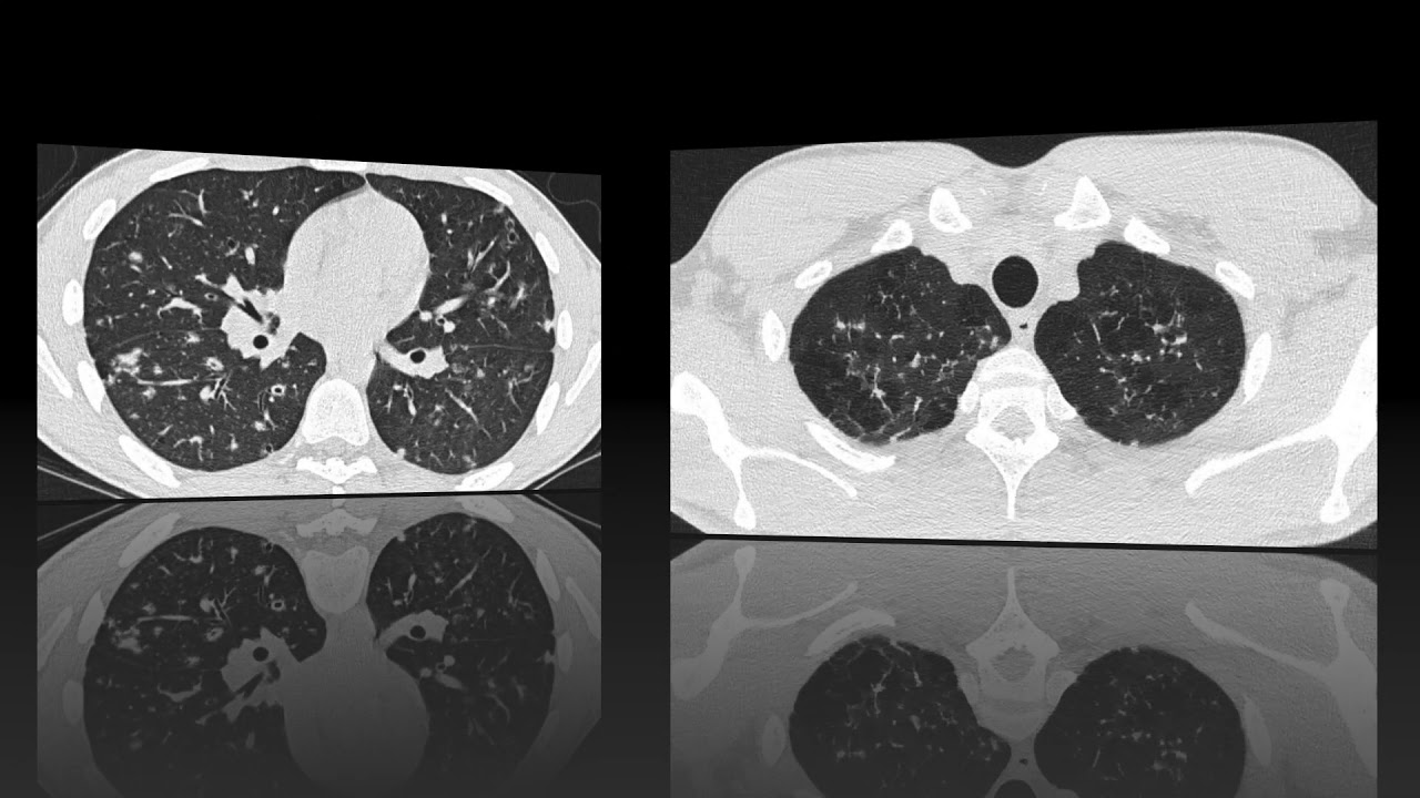
It consists of small centrilobular nodules of soft-tissue attenuation connected to multiple. These are due to filling of the.
Tree In Bud Sign Lungs
The connection to opacified or thickened branching structures extends.
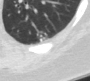
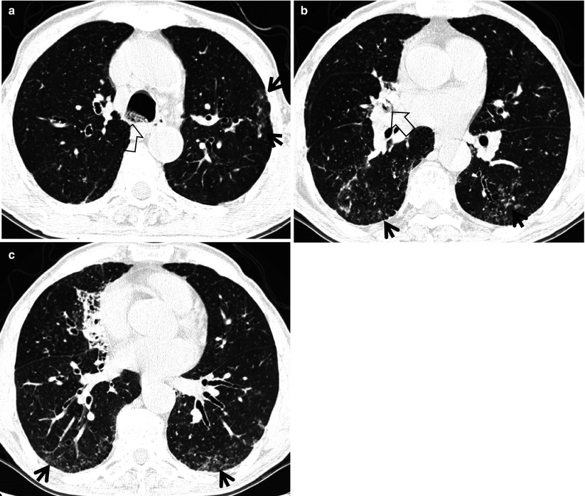
. Typically the centrilobular nodules are 2-4 mm in diameter and peripheral within 5 mm of the pleural surface. Tree-in-bud opacities appear as tiny centrilobular branching structures on CT most often in the lung periphery which resemble budding trees Figure 18-4. This abnormal growth is caused by a fungus that infects the trees bark.
The fungus causes the tree. 1 The tree-in-bud sign is a nonspecific imaging finding that implies impaction. Thus the bronchioles resemble a branching or budding tree.
Hi doctor My CT scan says defined streaky opacity with associated loss volume and clustered tree in bud nodules have developed in the anterior segment of the upper left. Although it was previously associated with. The tree-in-bud pattern can be seen on a thin-section computed tomography of the lungs.
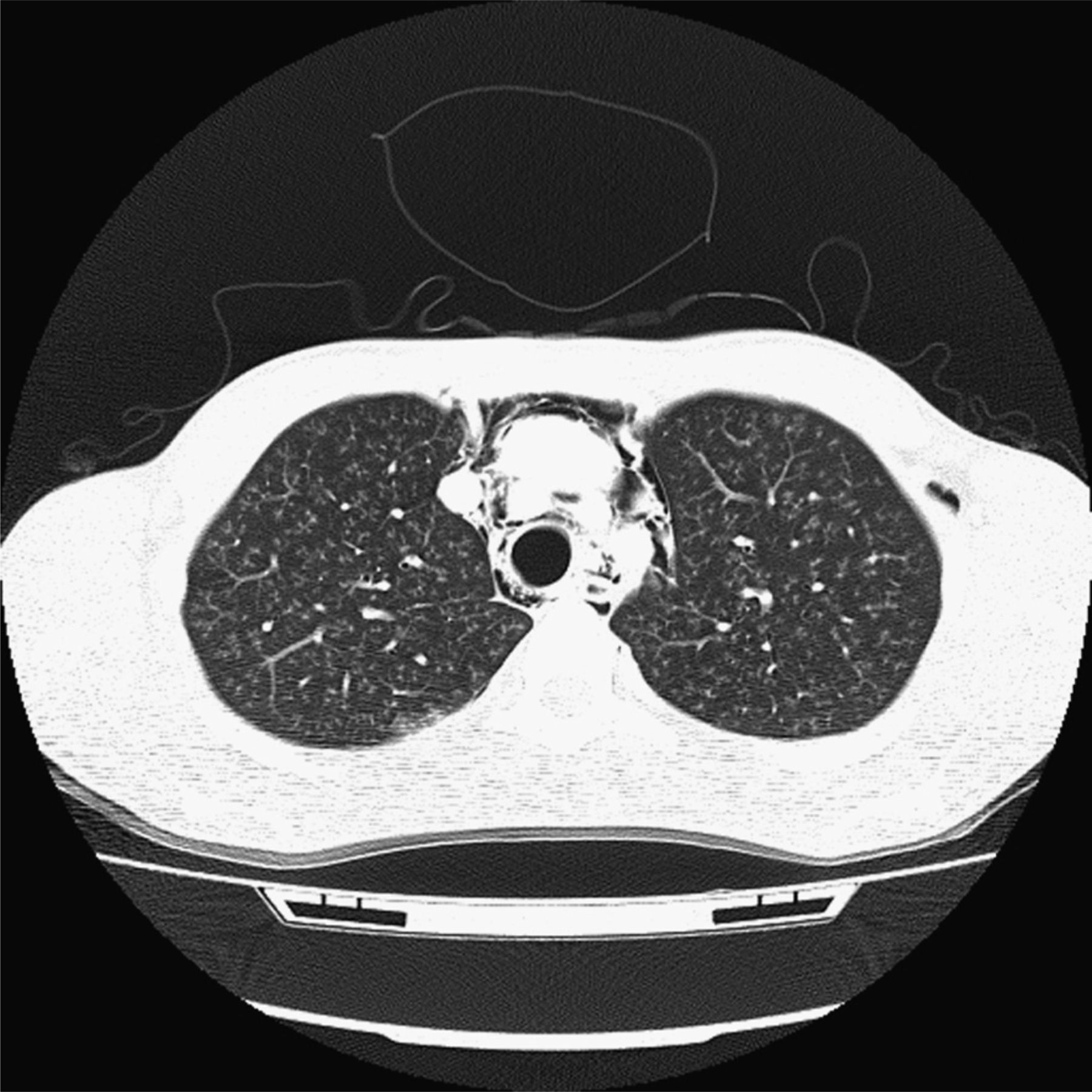
The tree in bud nodule is a growth that forms on the branches of certain trees. Tree-in-bud pattern seen on high-resolution CT HRCT indicates dilatation of bronchioles and their filling by mucus pus or fluid. Emboli can easily occur because tree-in-bud nodules most commonly represent active infection ie infectious bronchiolitis including among patients with underlying pulmonary metastases.
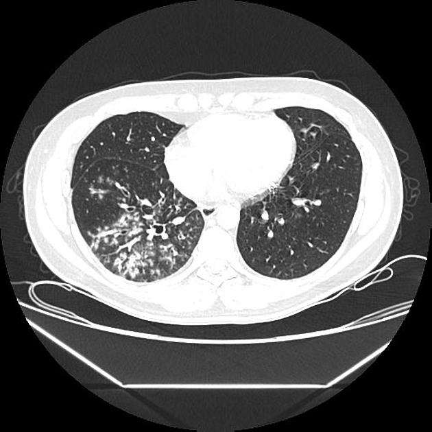
87 rows The tree-in-bud pattern is a special subset of centrilobular nodules initially described in CT scans of patients with endobronchial spread of Mycobacterium tuberculosis infection. Tree-in-bud sign refers to the condition in which small centrilobular nodules less than 10 mm in diameter are associated with centrilobular branching nodular structures 1 Fig. A tree-in-bud sign or pattern shows CT signs of multiple areas of centrilobular nodules with linear branching patterns.
In radiology the tree-in-bud sign is a finding on a CT scan that indicates some degree of airway obstruction. Tree-in-bud sign is not generally visible on plain radiographs 2It is usually visible on standard CT however it is best seen on HRCT chest.

Computed Tomography Of The Chest Showed Nodular Opacities With Tree In Download Scientific Diagram

Tree In Bud Pattern At Thin Section Ct Of The Lungs Radiologic Pathologic Overview Radiographics
Tree In Bud Attributable To Organising Pneumonia Thorax

Tree In Bud Pattern Radiology Case Radiopaedia Org

Tree In Bud Pattern Pulmonary Tb Eurorad

Tree In Bud Sign An Overview Sciencedirect Topics

High Resolution Computed Tomography Findings In Humoral Primary Immunodeficiencies And Correlation With Pulmonary Function Tests
Assessment Of Disease Activity And Complications In Patients Of Pulmonary Tuberculosis By High Resolution Computed Tomography Sudan Journal Of Medical Sciences Sjms

Co Rads 2 With Tree In Bud Sign A 27 Year Old Male Attended The Download Scientific Diagram

Tree In Bud Sign An Overview Sciencedirect Topics

Epos Trade

Tree In Bud Pattern At Thin Section Ct Of The Lungs Radiologic Pathologic Overview Radiographics
Tree In Bud Appearance Radiology Case Radiopaedia Org
The Radiology Assistant Hrct Common Diagnoses

Tree In Bud Pattern Pulmonary Tb Eurorad

Tree In Bud Pattern At Thin Section Ct Of The Lungs Radiologic Pathologic Overview Radiographics
Tree In Bud Sign Radiology Key

Pdf Tree In Bud Semantic Scholar
View Of Tree In Bud The Southwest Respiratory And Critical Care Chronicles